
Not sure what the difference between dental x-rays and CT-scans are? If you’re going in for a dental cleaning/exam, it’s good to know the difference beforehand, as they are both valuable tools in determining the health of your teeth and gums.
It’s also important to know if your dentist even offers CT-scan imaging. Many don’t, as it’s common (and dated) practice to only provide dental x-rays. Here at Perdido Bay Dental we offer X-rays, CT imaging, and other dental technologies, to ensure we’re getting the most accurate picture of each patient’s mouth.
What are Dental X-Rays?
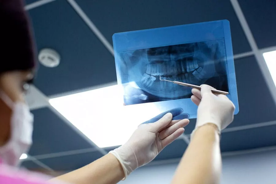
Dental X-rays are photographs of the teeth, bones, soft tissues and gums around them to help find problems and diseases within the teeth, mouth, and jaw. X-ray pictures can show cavities, teeth that have not yet erupted through the gums, and bone loss that cannot be seen during a standard visual examination. Dental X-rays may also be done as follow-up after dental treatments to make sure a procedure worked.
Types of Dental X-rays
- Bitewing X-rays show both the upper and lower back teeth in the same photo. These X-rays are used to show any decay in the teeth and how will the back teeth align when you bite down.
- Periapical X-rays show the entire tooth, from the exposed top of the tooth to the root and bone that supports the tooth. Periapical X-rays are used to find issues below the gumline that the naked eye cannot see, like cysts, bone loss, and impacted teeth.
- Occlusal X-rays show the top or bottom of the mouth, and help find potential extra teeth, or teeth that have not yet broken through the gum line. Occlusal X-rays also can show fractures in the jaw, cleft palate issues, and other growths.
- Panoramic X-rays show the entire lower jaw area, from the sinuses to the nose to the jaw. These X-rays can show any number of problems from bone abnormalities to cysts and growths to bite and jaw alignment issues.
What Are CT-Scans?
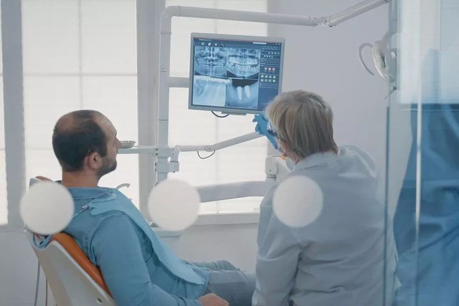
By contrast, a CT scan produces extremely detailed images of the body (or just mouth in this case) by taking a 360-degree image of an area. It can provide images of organs, bones, soft tissue, blood vessels and more, whereas an X-ray is primarily used to detect issues in bone. A CT scan is often used to diagnose cancer, musculoskeletal disorders, infections, and more. A dental CT scan creates a 3D image of dental structures, soft tissues, nerve pathways and bone in just one scan.
A traditional CT scan involves your entire body being placed into a large tube to create the images. A dental CT scan is a little different. It’s called a cone beam CT, where an X-ray beam in the shape of a cone is moved around the patient to create the images. A dental CT scan emits a lot less radiation than a traditional CT scan.
The dental CT scan helps to diagnose diseases of the jaw, bony structures of the face, nasal cavity and sinuses. However, it does not provide the full diagnostic abilities of the conventional CT, especially when it comes to muscles, lymph nodes, and nerves. Dentists use them to find impacted teeth, diagnose temporomandibular joint (TMJ) disorder, ensure accurate placement of dental implants, evaluating bone structure, and more.
Looking Deeper into Dental Imaging and Why It Matters
Dental appointments often move quickly. A short scan, a brief explanation, and then the next step begins. Still, that scan often guides every decision that follows. Dental imaging gives dentists a way to see beneath the surface, where many dental issues quietly begin. It supports better dental care by catching concerns early and helping protect long term oral health.
At Perdido Bay Dental, we do not just see imaging as some everyday thing. It gets woven into this whole careful setup where accuracy comes first along with making sure patients are comfortable and everything is explained clearly. When people learn why the imaging is needed it makes the whole dental visit seem less stressful, sort of more like it has a point.
Why Dental Imaging Matters More Than Many People Think
Teeth by themselves do not show everything that’s going on in the mouth. There are roots and bone levels and nerve pathways plus all the soft tissues that make things work right. Imaging is what lets dentists see those parts that stay hidden otherwise. Without that kind of help, problems just sit there until something like pain starts up or there is swelling.
It seems like even small things can be signs of bigger trouble. A little ache in your tooth or some tension in the jaw, or maybe sensitivity to hot or cold, those might mean deeper issues are brewing. Dentists use tools such as X-ray images to check it out, and there is also a cone beam CT scan for a closer look. Spotting stuff early with these makes a difference.
Traditional Dental X Ray and Its Everyday Value
Traditional X-rays remain a trusted part of modern dental technologies. They provide clear images of teeth and surrounding bones and work well for routine visits. Dentists often rely on traditional X-rays during cleanings and exams because they are fast and effective.
- These images help dentists:
- Find cavities between teeth
- Track bone loss over time
- Check the position of teeth below the gums
- Monitor previous dental work
Traditional dental X-ray images do have limits. They show flat views, which means some areas overlap. While this works for many situations, complex dental issues sometimes need more detail. That is where advanced imaging becomes helpful.
Understanding Cone Beam CT and Its Purpose
Cone beam CT, also known as cone beam computed tomography CBCT, provides a three-dimensional view of the mouth and jaw. Instead of a single flat image, this scan creates a detailed 3d image that shows depth and structure. Dentists can see how teeth, bone, and nerve pathways relate to one another.
Cone beam CT often supports treatment planning for:
- Dental implants
- Jaw alignment concerns
- Bone loss evaluation
- Impacted teeth
- Symptoms linked to TMJ disorder
The scan itself feels simple. Patients remain still while the machine rotates briefly. The result is a set of detailed images that guide safer and more precise care.
How Detailed Images Improve Treatment Planning
Treatment planning depends on clear information. When dentists understand what lies beneath the surface, they can choose options that fit each patient. Dental imaging removes guesswork and reduces surprises during procedures.
For dental implants, this matters greatly. Dentists must confirm enough bone exists and that nerve pathways remain protected. Cone beam CT makes these measurements possible. It also helps dentists plan placement angles and depth with confidence.
Detailed images also support follow-up care. Dentists compare scans over time to track healing and spot changes early. This ongoing insight keeps treatment focused and effective.
Dental Imaging and Comfort During Care
Imaging machines coming into sight usually trigger an uneasy feeling in most patients. This response can be considered reasonable. However, imaging makes it less likely to have to deal with discomfort later on. Early detection of problems gives the opportunity to dentists to deal with them before they become painful or complicated.
The case of jaw pain is a perfect example. A little stiffness may not be regarded as pressing even. The radiograph can point out the initial indications of a temporomandibular joint (TMJ) disorder, which will lead the dentist to recommend the use of supportive care. This scenario is the same for infections that are deep in the tissues and not visible on the surface. Imaging is the only method that can reveal such problems.
Dental imaging supports comfort by guiding thoughtful decisions. Patients benefit from fewer unexpected findings and clearer explanations.
How Dental Technologies Support Better Oral Health
The evolution of dental technologies is ongoing, but their main goal is still the same. They aid the dental practitioners in safeguarding dental health and achieving durable results. The imaging devices cooperate with the examinations and patient discussions to draw a complete picture of the dental health status.
The selection of imaging techniques is made according to the requirement by the dentists. Conventional X-rays are still advantageous for regular check-ups. The cone beam CT reveals more when problems with teeth or nerves or complicated structures dictate the treatment. Every tool shares the responsibility of good dental care.
Common Questions Patients Ask About Imaging
Many patients want reassurance before scans. Common questions often include:
How often imaging is needed?
Imaging is used only when it supports diagnosis or treatment planning, not at every visit.
Which scan fits their situation?
The dentist selects the scan based on the dental issue, symptoms, and level of detail required.
How images guide treatment choices?
Images reveal hidden problems and help dentists plan safer, more precise care.
Dentists explain these decisions clearly. Imaging is never used without purpose. Each scan supports accurate diagnosis and careful planning.
Clear Insight Leads to Confident Care
Dental imaging brings clarity to dental care. From traditional X-rays to cone beam CT, each option helps dentists see more and guess less. Patients gain confidence knowing their care rests on detailed images and thoughtful planning.
Understanding imaging removes fear and builds trust. When patients see how images guide treatment, visits feel more collaborative and less uncertain.
Schedule a Visit for Clear Answers and Thoughtful Care
Clear images lead to clear plans. Schedule a visit with Perdido Bay Dental to learn how dental imaging supports accurate diagnosis, careful treatment planning, and lasting oral health. A simple scan today can help protect comfort and confidence for years to come.
If you would like to schedule an appointment to have a dental CT-scan performed here at Perdido Bay Dental, please call us today at (850) 542-4428. We’re here to help.